Healing After Root Canal Microsurgery: Recovery Tips and Timeline
Healing After Root Canal Microsurgery: What to Expect
Root canal microsurgery, also known as an apicoectomy, is a precise surgical procedure performed at the tip of a tooth's root, typically when infection or inflammation has persisted after a previous root canal. Although the procedure itself is generally well tolerated, the days and months that follow involve a healing process that occurs in stages. Understanding what is normal during recovery, what is not, and how the body actually repairs the tissues involved makes the experience less unsettling and improves the likelihood of a smooth outcome. This article describes what to expect during recovery, the timeline over which healing occurs, and the steps that support the process.
What Happens to the Tissues During Recovery
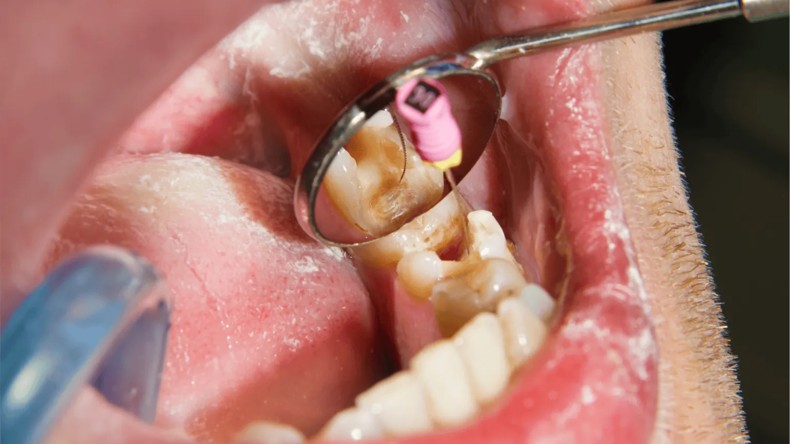
Microsurgery at the root tip involves several distinct tissues, each of which heals at a different pace. A small incision is made in the gum tissue to access the bone overlying the root, a small window is created in the bone to reach the root tip, the infected tissue is removed, the end of the root is sealed with a biocompatible material, and the gum tissue is repositioned and sutured. Healing therefore occurs across three layers: the gum tissue at the surface, the bone surrounding the root, and the smaller blood vessels and connective tissue between them.
The gum tissue heals most quickly. Within a few days, the incision begins to close, and within one to two weeks the surface tissue has substantially recovered. The bone surrounding the root takes much longer. Bone is a living tissue that remodels gradually, and the defect created during surgery, along with any area of bone loss that existed before surgery as a result of infection, fills in over months rather than weeks. This is why the tooth can feel slightly different on biting for some time after the procedure, even when the surface tissue has fully healed, and why follow-up radiographs are taken at intervals over the first year to confirm that the bone is responding as expected.
The First 24 to 48 Hours
The first two days after surgery are when discomfort, swelling, and any minor bruising are most noticeable. Swelling typically reaches its peak on the second day rather than the first and then begins to subside. Mild bleeding or oozing from the surgical site for the first several hours is normal and is managed by applying gentle pressure with clean gauze. A cold compress applied to the outside of the face for short intervals during the first day reduces swelling and provides some relief. Pain is generally managed with the medication prescribed or recommended by the treating endodontist, most often an anti-inflammatory such as ibuprofen, sometimes combined with a stronger medication for the first day or two when the surgical site was more extensive.
During this period the patient is generally advised to rest, to keep the head slightly elevated when lying down, to avoid rinsing or spitting vigorously, and to avoid disturbing the surgical site with the tongue or with a toothbrush. Soft foods at moderate temperatures are easier to manage than hot or cold foods, and chewing should be directed to the opposite side of the mouth.
The First Week
Between the second and seventh days after surgery, the initial swelling subsides, the surface tissue begins to close, and most patients notice a clear improvement in how the area feels. Sutures, if not the self-dissolving type, are typically removed at a follow-up visit during this week. The patient is generally able to return to most normal daily activities within a day or two of the procedure, although strenuous physical activity is best deferred for several days, since increased blood pressure during exercise can disturb the surgical site and prolong recovery.
Oral hygiene around the surgical site requires care. The teeth elsewhere in the mouth should continue to be brushed and flossed normally, but the area near the incision is best left undisturbed for the first few days, after which gentle brushing can be resumed. A warm saltwater rinse, used several times a day starting the day after surgery, helps to keep the area clean without disturbing the healing tissue. Patients who smoke are advised to refrain for as long as possible during this period, since smoking significantly impairs the healing of both gum tissue and bone.
Weeks Two Through Six
By the second week, the surface tissue has substantially healed, and most patients have returned to a normal diet, although chewing very hard or crunchy foods directly over the surgical site is best avoided for a longer period. The tooth itself may continue to feel slightly different on biting, since the surrounding bone is still in the early stages of remodeling, and some patients describe a sensation of the tooth feeling slightly loose. This sensation is generally normal during this period and resolves as the bone fills in around the root.
A follow-up visit during this period allows the treating endodontist to confirm that the gum tissue has healed as expected and that there are no signs of persistent infection. In most cases, no further intervention is required at this stage, and the next appointment is scheduled several months later for radiographic follow-up.
Bone Healing Over the Following Months
Bone regeneration is the slowest part of recovery and continues well after the patient feels fully healed. The bone defect at the root tip, including any area that had been resorbed as a result of the infection that led to surgery in the first place, gradually fills in with new bone over a period that typically ranges from six months to a year or longer in more extensive cases. Follow-up radiographs taken at six and twelve months allow the progress of bone healing to be confirmed, and the appearance of the surrounding bone is one of the key indicators used to determine whether the surgery has succeeded.
The success rates reported in the published literature for modern root-end microsurgery are high. Studies using current surgical techniques, magnification, and biocompatible root-end filling materials commonly report success rates in the range of 90 percent or higher at follow-up intervals of five years or more, which is one of the reasons surgery is increasingly chosen over extraction when conventional retreatment is not an option.
What Is Normal and What Is Not
Mild to moderate discomfort, swelling that peaks on the second day and then improves, minor bruising of the cheek or skin near the jaw, mild oozing from the site during the first day, and a sensation that the tooth feels slightly different on biting are all expected findings during recovery and do not require additional intervention.
Findings that should prompt a call to the treating endodontist include swelling that worsens after the third day rather than improving, increasing rather than decreasing pain over the days following surgery, fever, drainage of pus from the site, persistent bleeding that does not respond to pressure, or numbness in the lip or chin that does not resolve in the days following surgery. These findings are uncommon but warrant prompt evaluation when they occur, since early intervention generally produces a better outcome than waiting.
Supporting Recovery
Several straightforward measures support the healing process. Resting during the first day, applying cold compresses during the first day and switching to warm compresses after the first 24 hours if mild swelling persists, taking prescribed or recommended medications on schedule, completing the full course of any prescribed antibiotic, eating soft foods at moderate temperatures, and avoiding tobacco use are the measures most directly supported by the evidence. Maintaining good oral hygiene elsewhere in the mouth while protecting the surgical site, attending scheduled follow-up appointments, and reporting any concerning findings rather than waiting until the next scheduled visit complete the picture.
Most patients are surprised at how manageable recovery from an apicoectomy actually is once they understand what is happening at each stage. The early discomfort is usually milder and shorter than expected, and the longer process of bone healing occurs in the background while the patient resumes normal life.
About Tri-City Endodontics
Dr. Malhan and the Tri-City Endodontics team have practiced in Pasco for more than 25 years and perform root-end microsurgery for cases referred from general dentists throughout the Tri-Cities region. Each surgical case is evaluated with three-dimensional imaging to assess the position of the root, the extent of the lesion, and the proximity of anatomical structures such as the sinus or nerve canals, and patients are informed of these findings and the realistic prognosis before surgery is scheduled. Follow-up visits and radiographs over the months and years after surgery are scheduled to confirm that healing is progressing as expected, so any concern can be identified and addressed promptly