How 3D Imaging Is Revolutionizing Root Canal Diagnosis and Treatment
When Your Endodontist Recommends a CBCT Scan: What You Should Know
Patients referred to an endodontist for root canal evaluation are sometimes told that a cone beam CT scan, or CBCT, is recommended in addition to or instead of conventional dental X-rays. The recommendation can prompt reasonable questions: Why is the additional scan needed? Is it always necessary? How much radiation is involved? What will the scan actually show that a standard X-ray cannot? This article addresses those questions from the patient's perspective, focusing on how the decision to use CBCT is made and what to expect when it is recommended.
Why an Endodontist May Recommend a CBCT Scan
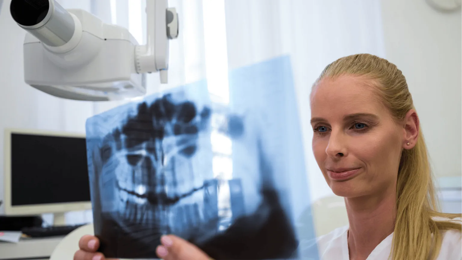
A conventional dental X-ray, called a periapical radiograph, captures the tooth and surrounding bone as a single two-dimensional image. The image is useful for many diagnostic questions and is the appropriate first step in most endodontic evaluations. The limitation is that structures lying along the path of the X-ray beam appear superimposed on a single image, which means that small or subtle features can be obscured by surrounding bone or by the roots of adjacent teeth.
CBCT addresses this limitation by capturing a volume of tissue that can be examined as a series of thin slices from any angle. For specific clinical questions, the three-dimensional information provides answers that a conventional X-ray cannot. The decision to recommend a CBCT scan is generally driven by one of these questions arising during the evaluation.
The most common reasons for the recommendation include suspicion that the canal anatomy of the tooth is more complex than a conventional radiograph can fully evaluate, a previous root canal that has failed and whose cause needs to be identified before retreatment is considered, suspected cracks or fractures in the root that cannot be confirmed on two-dimensional imaging, suspected resorptive defects in which the tooth structure is being broken down from within, and planning for surgical procedures where the proximity of anatomical structures such as the maxillary sinus or the nerve canal in the lower jaw is relevant.
If the clinical situation does not raise any of these questions, a CBCT scan is generally not necessary, and a standard radiograph alone provides the information needed for the case.
What the Scan Will Actually Show
For a patient who has been told that a CBCT is recommended, knowing what the scan will show beyond what a conventional X-ray can provide helps make the conversation about findings more meaningful.
Additional canals in the tooth that may have been missed in previous treatment or that need to be addressed in planned treatment are often visible on CBCT when they cannot be clearly seen on conventional X-rays. The MB2 canal in upper molars, which is present in most of these teeth but is often missed in conventional root canal treatment, is a frequently cited example. The exact shape, curvature, and length of each root, which affect how a procedure will be performed and what instruments will be used, can be assessed in three dimensions rather than estimated from a two-dimensional projection.
Periapical lesions, the areas of bone loss that develop around the tip of an infected root, are often visible on CBCT before they are large enough to be apparent on conventional radiographs. This is significant because the appearance and size of these lesions is one of the main indicators used to confirm that endodontic treatment is needed and to monitor healing afterward. In a previously treated tooth that is symptomatic, identifying a periapical lesion that is not yet visible on conventional radiographs can change the diagnosis and the recommended treatment.
Resorptive defects, in which the body's own cells are breaking down a portion of the tooth structure, can be located precisely on CBCT in a way that determines whether the defect can be treated and what approach is appropriate. The position of the root tip in relation to the maxillary sinus or to the nerve canal in the lower jaw, important for planning surgical procedures, is visible on CBCT in a way that supports safer and more predictable surgery.
Radiation Considerations
A CBCT scan involves more radiation than a conventional dental X-ray, although less than a medical CT scan covering the same area. The actual dose depends significantly on the scan parameters, including the size of the area being imaged and the resolution settings selected. Modern endodontic CBCT protocols are designed to limit the field of view to the specific tooth or small region being evaluated, which substantially reduces the radiation dose compared with larger scans covering the whole jaw or head.
The principle that guides the use of CBCT, supported by the published guidelines of professional endodontic organizations, is that imaging should be selected based on the specific diagnostic question being asked and should provide information that cannot be obtained from less radiation-intensive alternatives. The scan is appropriate when it will change the diagnosis or the treatment plan and is not appropriate as a routine examination for every case.
A patient who is unsure whether the additional scan is necessary in their specific case can reasonably ask what question the scan is intended to answer and whether the answer would change the recommended treatment. A clinician who has recommended CBCT for a clear reason can explain the reason; if no clear reason can be articulated, the scan may not be necessary.
What the Experience Is Like
A dental CBCT scan is brief and straightforward. The patient stands or sits in the imaging unit, holds still while the unit rotates around the head, and the scan completes in less than thirty seconds. There is no injection, no contrast material, and no sensation during the scan itself. The patient receives the same lead apron and thyroid collar used for conventional dental X-rays when appropriate to the scan being performed.
The resulting images are available for review within minutes. Many endodontists review the images with the patient on a screen in the office, rotating the volume to show specific findings, and explain how the findings affect the diagnosis and the recommended treatment. This conversation is often more concrete than a discussion based on a conventional X-ray, since the three-dimensional images make features such as canal anatomy and bone loss more visible to a patient who is not trained to read radiographs.
Questions a Patient Can Ask
A few specific questions allow a patient to understand whether the CBCT is appropriate in their case and what will come of it.
What clinical question is the scan intended to answer is a useful starting point. A clear answer indicates that the recommendation is driven by a specific concern. A vague answer may indicate that the scan is being recommended routinely rather than for a specific reason.
How the findings would change the treatment is the natural follow-up. If the answer to the clinical question would change what is recommended for the tooth, the scan is providing information that affects the decision. If the answer would not change the recommended treatment, the scan may not be necessary.
Whether the scan will be limited to the area of the tooth in question rather than covering a larger area is worth confirming. Smaller field-of-view scans involve less radiation and produce higher-resolution images of the specific area being evaluated.
What the findings actually showed, after the scan, is worth discussing. The patient has a reasonable interest in understanding what was identified, since the findings often affect the prognosis and the recommended approach.
When CBCT Is Not Necessary
For most routine endodontic cases, a CBCT scan adds little to the diagnosis or the treatment plan. A straightforward root canal on a tooth with simple anatomy and no unusual findings on conventional radiographs generally does not require three-dimensional imaging. A clinician who recommends CBCT for every case, regardless of complexity, is not following the imaging principle that guides current practice, and a patient is reasonable to ask why the additional scan is needed in their specific situation.
The most useful framing is that CBCT is a powerful tool for specific situations rather than a routine part of every endodontic evaluation. When the clinical situation raises a question that three-dimensional imaging can answer, the scan provides information that improves the diagnosis and the treatment plan. When the situation does not raise such a question, conventional imaging is generally sufficient.
About Tri-City Endodontics
Dr. Malhan and the Tri-City Endodontics team have practiced in Pasco for more than 25 years and use CBCT imaging as part of the diagnostic and treatment planning process for cases referred from general dentists throughout the Tri-Cities region. The decision to obtain a CBCT scan is made on a case-by-case basis, with scans performed at the smallest field of view consistent with answering the clinical question. Findings from the scan are reviewed with the patient and the referring dentist when relevant, so the additional information contributes to a clear understanding of the condition of the tooth and the realistic prognosis of the treatment options under consideration