How Dental Microscopes Improve Treatment Success
What a Dental Microscope Means for the Patient During Root Canal Treatment
Patients arriving for root canal treatment in a specialist endodontic office often notice the dental operating microscope as the most visually distinctive piece of equipment in the room. The microscope has become standard in specialist endodontic practice over the past two decades, and its presence affects what the clinician sees during the procedure, how certain steps are performed, and how the findings can be communicated to the patient. This article describes what the microscope means from the patient's perspective: what changes about the appointment, what kinds of cases benefit most, what to ask about, and what the technology does and does not change.
What the Microscope Actually Does
The dental operating microscope provides two things that cannot be obtained from other equipment in the dental office. The first is high magnification of the working area, generally in the range of four to twenty-five times depending on the setting selected during the procedure. The second is coaxial illumination, which lights the field of view directly along the same axis as the operator's line of sight. The combination is what allows the operator to see clearly into a canal that is less than a millimeter in diameter and several millimeters deep.
For context, the structures the operator works with during root canal treatment are genuinely small. The opening to an additional canal that needs to be located in an upper molar may be smaller than the head of a pin. Cracks that determine whether a tooth can be saved or needs to be extracted may be barely visible features on the floor of the pulp chamber. The precise location at which to seal the end of a root during apicoectomy is a few millimeters of canal anatomy that needs to be evaluated and prepared accurately. These are the kinds of features that the microscope is designed to address.
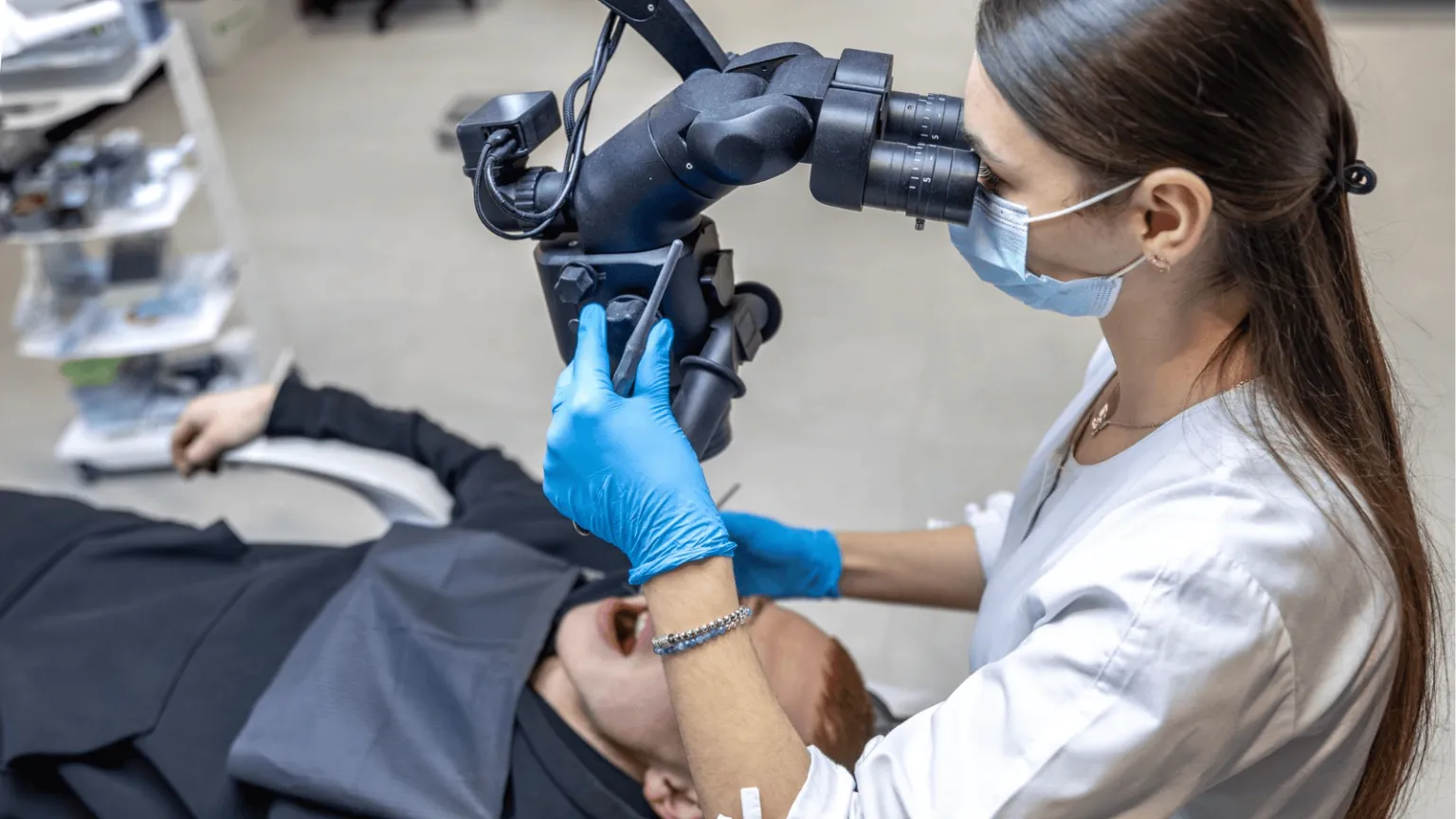
What the Patient Notices During the Appointment
From the patient's perspective, a procedure performed under the microscope is not substantially different from a procedure performed without it. The operator sits slightly behind the patient and works through the eyepieces of the microscope rather than leaning over the patient directly, which can actually be more comfortable for the patient than having someone working very close to their face for an extended period. The microscope itself is positioned above and behind the patient and does not contact them at any point.
The procedure may take slightly longer in complex cases because the operator is taking more time to inspect the anatomy carefully at each step rather than relying on tactile feedback alone. In simpler cases, the time difference is minimal. The patient experiences the same sequence of steps that any root canal involves: local anesthesia, placement of the rubber dam, access to the canal system, cleaning and shaping, sealing, and a final restoration.
Many practices using the microscope record still images or video during the procedure. These can be reviewed with the patient afterward to show the actual interior of the tooth, the canal anatomy that was located, and any unusual features that were identified. This is often the first time a patient sees what the inside of their own tooth looks like, and it tends to make conversations about prognosis, follow-up, and any remaining concerns more concrete. A patient who has seen the crack on the floor of the pulp chamber that explains why an earlier root canal failed has a clearer understanding of why retreatment may not be the right next step than a patient who has only been told about it.
What Patients Should Know About When the Microscope Matters Most
Not every endodontic case requires microscopic visualization to produce a good outcome. A straightforward root canal on a tooth with simple anatomy and no prior treatment can produce a good result without high magnification. The microscope contributes most clearly in specific situations, which is useful information for a patient considering treatment in a more complex case.
Retreatment of a previously failed root canal is the situation where the microscope's contribution is most apparent. Identifying why the original treatment failed often depends on finding a missed canal, a crack, a perforation, or a separated instrument, and these findings can be difficult to make reliably without magnification. The information from this examination determines whether retreatment is the right approach or whether another option such as apicoectomy or extraction would have a better prognosis.
Surgical endodontics, including apicoectomy, depends on the microscope for direct visualization of the root tip during preparation and sealing. Studies comparing modern microscopic apicoectomy to older surgical techniques show substantially higher success rates for the microscopic approach, and the equipment is now considered essential for this type of surgery rather than optional.
Teeth with complex anatomy, including upper molars with multiple canals, teeth with calcified canals where age or chronic inflammation has narrowed the canal space, and teeth with unusual root curvature, generally benefit from microscopic visualization. Suspected fractures, resorptive defects, and perforations from previous dental procedures are all situations in which the microscope contributes meaningfully to the diagnosis.
For these cases, the microscope is not a marketing feature. It is part of how the case is evaluated and treated, and patients with complex situations are reasonable to ask whether it will be used.
Questions a Patient Can Ask
A few specific questions allow a patient to understand what role the microscope will play in their treatment.
Whether the microscope will be used throughout the procedure or only at selected steps is a useful question. Some practices use the microscope continuously, while others use it primarily for specific steps. Either approach can produce good results, but the answer indicates how the clinician thinks about the equipment.
Whether the practice can provide images or video from the procedure is another reasonable question. Many specialist practices routinely do this, and the documentation is useful both for the patient's understanding and for any future treatment or second opinion.
What the clinician's specific training and experience are with the procedure being considered is the most important question. The microscope is a tool, and the outcome of any procedure depends on the clinician using it. A practice that has been using the microscope for years on cases similar to the patient's will generally produce a more predictable result than a practice that has recently added the equipment.
What the Microscope Does Not Change
The microscope improves what the clinician can see, but it does not by itself determine the outcome of treatment. The result of a root canal still depends on accurate diagnosis, careful technique, proper disinfection of the canal system, a good final seal, and timely placement of a permanent restoration after the canals are sealed. A tooth with a vertical root fracture cannot be saved by any technique. A tooth where the final restoration is delayed for months will eventually develop problems regardless of how carefully the canal system was treated. A tooth in a patient with significant medical conditions that affect healing will respond differently from a tooth in a patient without such conditions.
This is worth stating directly because the marketing of dental technology can leave the impression that a single piece of equipment guarantees success. In reality, the microscope is one component of a procedure that depends on several factors working together. The most useful framing for a patient is that the microscope improves the predictability of complex cases, makes findings that would otherwise be missed visible, and supports clearer communication about what is happening inside the tooth, all of which contribute to a better-informed decision and a better-executed procedure, without being responsible for the outcome on its own.
About Tri-City Endodontics
Dr. Malhan and the Tri-City Endodontics team have practiced in Pasco for more than 25 years and use a dental operating microscope as part of standard practice for root canal therapy, retreatment, and surgical endodontics on cases referred from general dentists throughout the Tri-Cities region. Findings observed under magnification, including features that cannot be seen on conventional radiographs or with the unaided eye, are reviewed with the patient and the referring dentist when relevant, so that decisions about treatment are made with a clear understanding of the actual condition of the tooth